Assisted reproduction
Seminogram: sperm testing and evaluation
A seminogram, also called a spermiogram or sperm analysis, is the diagnostic test most commonly used by assisted reproduction centres to assess male fertility.
This technique consists of qualitatively and quantitatively analysing a sperm sample at the macroscopic and microscopic levels. Additionally, a seminogram is used as a screening test after performing a vasectomy so as to ensure that the procedure was effective.
How should the sample for a seminogram be obtained?

In order to guarantee the maximum reliability of the seminogram or spermiogram diagnosis, the sperm sample must be obtained following the guidelines below:
- Maintain sexual abstinence for 2 to 4 days. The limits are no less than 2 days and no more than 4 days of abstinence.
- Gather the sample in a sterile container: Ginefiv will provide one but they are also available for purchase in any pharmacy.
- The way that the sample is obtained must be masturbation, and neither coitus interruptus nor the use of commercially available condoms is valid.
- Maintain maximum hygiene conditions for the sample collection (wash hands beforehand, etc.).
- The sperm sample must be complete; if any is spilled, the sample will not be valid. If you have any doubts, our biologists will tell you whether or not the test can be performed.
If the sample is collected at home:
- Once the sample has been obtained, you must ensure that the container is fully closed and that its temperature does not drop (it must be kept at body temperature [36°-37°], avoiding brusque temperature changes) and therefore we recommend carrying it in an inner pocket.
- The maximum amount of time between collection and delivery to the lab must be less than an hour and a half. If you have had a fever in the 14 days prior to the collection of the sperm sample or if you have taken any antibiotics, you must contact the lab to postpone the appointment
What aspects are evaluated in the seminogram or spermiogram?
The WHO established a series of parameters when performing a semen analysis and within them we can differentiate between macroscopic and microscopic parameters (WHO 2021).
The macroscopic parameters under study in a seminogram are:
- Appearance: The appearance of the semen is evaluated in terms of its colour, opacity or transparency, and the presence of gelatinous elements. Human sperm is usually a homogenous, opalescent, and whitish-grey liquid with a yellowish tint. Anomalies in its appearance can show the presence of abnormal compounds in the ejaculate (e.g. haematospermia: when there are blood cells in the sample, giving it a pinkish tinge), as well as the variation and concentration of certain ejaculate components.

- Liquefaction: This occurs when the sample is left to settle for 20 minutes at room temperature; the semen become less compact and the sample can then be analysed correctly. When liquefaction takes longer than 60 minutes, this is considered abnormal and must be indicated in the report.
- pH: The pH value measures the acidity or alkalinity of the sample, with the normal value being equal to or above 7.2. Given that the secretions of the seminal vesicles are alkaline, a variation of the sample’s pH could indicate certain disorders.
- Volume: This is measured in ml. The normal volume of semen in a humanejaculation is over 1.4 ml and under 6 ml. This volume is the result of thesecretions of the prostate, seminal vesicles, epididymis, and the testicles (only10% of the total composition of the ejaculate). The volume of the ejaculate canvary based on prior sexual abstinence as well as sexual excitation. In certainpathologies, the volume of the semen sample may be anomalous, either aboveor below the range which is considered typical. Cases in which the volume islower than 1.4 ml are called hypospermia and the term hyperspermia is usedwhen the volume is over 6 ml.
- Colour: semen should have a pearly colour. If it is slightly transparent, this could be due to the presence of leukocytes (white blood cells) in the ejaculate.
The main microscopic parameters that are evaluated in a seminogram are as follows:
Sperm concentration:
The number of spermatozoa per millilitre of sperm, generally measured in millions per millilitre. The spermiogram report must show the concentration per millilitre and the total concentration in the ejaculate. A sample is considered normal when the concentration of spermatozoa per millilitre is over 16 million and the concentration of the entire ejaculate is over 39 million. Values below these parameters determine what is known as oligozoospermia.
Sperm motility:
The movement capacity of the spermatozoa is analysed, checking for distinct motility patterns:
- PR or progressive motility.
- NP or non-progressive motility (“in situ” movement, with no progression).
- IM or immotile: spermatozoa with no movement.
A sample is deemed to have a normal motility parameter when the PR value is over 30% and the sum of PR+NP is 42% (both requirements are necessary). When these requirements are not fulfilled, the term used is asthenozoospermia.
Vitality: Sperm vitality is reflected in the percentage of spermatozoa which are alive. This parameter does not always necessarily coincide with the percentage of immotile spermatozoa. The normal value is when the percentage of living spermatozoa is over 54%. When it is below this value, it is called necrozoospermia
Morphology:
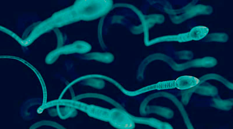
This parameter refers to the shape of the spermatozoon. A spermatozoon is composed of three parts: the head, the neck or middle piece, and the tail. There are various criteria for the determination of the morphology of a spermatozoon and each one of these establishes a cut-off point for the determination of what is normal. The WHO (2010) recommends the use of the strict Tygerberg criteria, which deems a sample to be normal when at least 4% of the spermatozoa have the normal morphology according to its own criteria. If the number is below this value, it is called teratozoospermia.
The presence of agglutination:
The agglutination of motile spermatozoa is when a number of them are joined together. They can be stuck together by their heads, by their tails or head-to-tail. The presence of agglutinations may suggest infertility of immunological origin. If agglutination is observed in a basic spermiogram, then other, more specific tests need to be performed to rule out this possible immunological disorder.
The presence of other cellular components and detritus:
Sperm normally contains cells other than spermatozoa, such as epithelial cells, spermatogenetic cells and leukocytes. The excessive presence of other types of cells can indicate certain anomalies or pathologies. In the case of leukocytes, the presence of less than one million per ml is considered normal. If a greater concentration is found, we use the term leukocytospermia.
What conclusions can be drawn from a seminogram or spermiogram?
Studying the basic parameters described above can determine to a certain extent the functionality of the male sexual glands and can therefore guide us in the identification of certain pathologies or in predicting the male’s fertility. In certain situations, we may need a more in-depth study of other parameters that are not taken into consideration in a basic sperm analysis or spermiogram
Other terms of interest related to seminograms
Aspermia: a total absence of ejaculate. On some occasions this is due to retrograde ejaculations (when the sperm passes to the urinary bladder after a sexual act).
Azoospermia: the total absence of spermatozoa in the ejaculate. When the ejaculate is diagnosed with azoospermia, this does not mean that spermatozoa are not produced in the testicles, only that they are not present in the ejaculate. There are different types of azoospermia and they can occur due to different causes. Therefore, if the diagnosis of a spermiogram is azoospermia, then a more in-depth study of the male needs to be carried out.
Cryptozoospermia: this is the presence of a very low concentration of spermatozoa in the ejaculate.


